Multiverse Journal - Index Number 2215: 21st May 2025, Some Background, Example of Basic Medical Science Research Results
A Journal across Realities, Time, Space, Soul-States.
—
May 21st, 2025
Good Tuesday,
May the Spirit of the Gospel and the Holy Word be Always on our Tongues, in our Hearts, Minds, and in our Hands.
Holy Virgin Mother Mary and All Saints - Pray for us!
—
Index Number 2215:
— —
—
May this article find us all ever closer to God, and His Clarity.
Example of my basic medical research results for background information. This paper used my raw image Capture, image data analysis, creating figures, and as per last author was involved significantly in document content. ..
.. and Bla-Bla-Bla .. I was Kiss My Ass excellent at this type of work I shaped myself to be from 5th grade and during this earlier period of work when it was still a ‘Dream Job’ and my Joy-Love-Hope is my life was not murdered by the Hell of the vile Witch, her coven and delusional psychotic [white Christian virtue-based raised] man hating Meritless Feminists that turn all spaces they sicken into Toxic hell-holes because of resentment, sniff of decent masculine affect & Works, .., but perhaps mostly when (and not if, but always) we fail to worship them for their Magic-Genitals.
So, .. Fuck You, Synagogue of Satan, and your Witches and Minions.
God Bless., Steve
Paper Citation: David M. Warshaw et al., Corkscrew-Like Shortening in Single Smooth Muscle Cells. Science 236, 1457-1459(1987). DOI:10.1126/science.3109034
2nd version of an AI generated audio overview conversation:
URL: https://notebooklm.google.com/notebook/4a192265-ca60-47c3-9acd-fc959af023e3
Corkscrew-Like Shortening in Single Smooth Muscle Cells
David M. Warshaw*, Whitney J. McBride, Steven S. Work Department of Physiology and Biophysics, University of Vermont, Burlington, VT 05405
*To whom correspondence should be addressed.
Abstract: The unique corkscrew-like shortening of single smooth muscle cells, with a pitch of 1.4 cell lengths at a rate of 27 degrees per second, was observed using digital video microscopy to track marker bead movements on the cell surface. This shortening mechanism suggests a helical arrangement of the contractile apparatus or cytoskeleton (or both) within the cell, which may contribute to the characteristic slower and more economical contraction of smooth muscle compared to skeletal muscle.
Smooth muscle contracts more slowly and economically than skeletal muscle, possibly due to the organization of its contractile apparatus. The way a single smooth muscle cell shortens is determined by the arrangement of its contractile apparatus. The shortening of contracting cells was studied by tracking marker bead movements on the cell surface using digital video microscopy. Smooth muscle cells were observed to shorten freely in a distinct corkscrew-like manner, with a pitch of 1.4 cell lengths (the length change needed for one full rotation) at a rate of 27 degrees per second. This corkscrew-like shortening was interpreted using a structural model where the contractile apparatus or cytoskeleton (or both) are helically oriented within the cell. This arrangement of cellular architectural elements may partly explain the contractile capabilities of smooth muscle.
The ability of smooth muscle to actively shorten its length results from the cyclic interaction of myosin cross-bridges with actin, generating force to slide neighboring actin filaments past myosin filaments. However, the arrangement of these filaments into specific contractile units and their orientation within smooth muscle cells are not well-defined. Smooth muscle contraction is characterized by a slower shortening velocity and the ability to generate comparable or greater force per cross-sectional area with far less myosin than skeletal muscle. Therefore, understanding contractile unit arrangement and its interaction with the cytoskeleton in smooth muscle could help explain these differences in contractile properties between the two muscle types.
Structural models have been proposed for the arrangement of contractile units within smooth muscle cells. Most models share a common feature, first described by Rosenbluth, where contractile units attach to the cell membrane and traverse the cell interior at small angles to the cell's long axis. More recently, Small and Fisher and Bagby proposed that contractile units are attached helically along the cell membrane. If this proposal is correct, an isolated single cell, fixed at one end and allowed to shorten freely, would be expected to do so in a corkscrew-like manner. To test this hypothesis, single smooth muscle cells were enzymatically isolated from the stomach muscularis of the toad Bufo marinus, and their structural changes during free shortening were visually monitored.
Materials and Methods
To characterize the shortening motion of isolated smooth muscle cells, a low calcium (0.18 mM) physiological salt solution containing 105 cells per milliliter was mixed with a similar cell-free solution containing positively charged anionic-exchange resin beads, 1 µm in diameter (9 x 10⁶ per milliliter). These charged beads strongly adhered to the cell membrane, providing numerous visual markers to decorate the cell surface (Fig. 1). A single smooth muscle cell was then electrostatically attached at one end to a larger, 20-µm anionic-exchange resin bead, which was glued to the end of a micropipette, under a microscope using a micromanipulator (Fig. 1). To ensure the entire cell length was in focus, a second micropipette, without a resin bead, was attached to a micromanipulator and used to lift the cell into the focal plane. The cell was then electrically stimulated to initiate contraction and induce cell shortening. As the cell shortened, its image was recorded on videotape and replayed through an IBM PC-XT laboratory computer, where single frames were digitized every 400 msec. The microscope objective's depth of field (8.0 µm) allowed bead images to be distinguished on all cell surfaces.
Our results show that, as predicted by the Small and Fisher and Bagby models, single smooth muscle cells shorten in a corkscrew-like manner. The cell shortened by 30% of its initial length during the first 4.8 seconds after electrical stimulation, at an average rate of 0.11 cell length per second (Fig. 1), a rate similar to previously reported values. However, when marker bead images were tracked, it was evident that their position on the cell surface changed during cell shortening (Fig. 1).
To more precisely define single-cell shortening by monitoring changes in bead position, a computer-based analysis of successive digitized images was developed to determine: (i) cell length, (ii) relative position along the cell length where a bead was located, (iii) cell diameter at a bead location, and (iv) angular bead position on the cell circumference.
The method of cell attachment to the microprobe and potential non-uniformities of contraction after activation were investigated as possible explanations for the observed corkscrew-like shortening. To control for the attachment mode, decorated cells resting on a glass slide were also observed to rotate during contraction. Regarding non-uniformities in activation, the relative bead position along the cell length as a function of the percentage of cell shortening (Fig. 2A) indicated no significant shifts in relative bead positions during shortening. Thus, for a bead to maintain a constant relative position along the cell length as it shortened, uniform cell shortening must have occurred. While the relative position of the bead along the cell length remained constant during shortening, significant changes in the angular position of a bead on the cell surface were observed, indicating that the cell rotated as it shortened. In the 15 cells studied to date, there appears to be no preferential direction of rotation.
From the data on the change in angular position of a bead, it appeared that beads at different relative positions along the cell length rotated at varying speeds after activation (shown as different slopes in Fig. 2B), and that the maximum rate of angular change increased the farther the bead was from the fixed cell end (Figs. 1C and 2D). These changes in bead angular position were also used to describe the corkscrew-like shortening of the cell in terms of a helical pitch. An estimate of bead pitch was obtained from the slope of the relationship between the angular change of a bead and the percentage of cell shortening after activation (Fig. 2C). For the cell in Fig. 1, a bead pitch of 1.4±0.1 (n = 7) cell lengths was obtained during the initial 20% of cell shortening.
After contraction, cells spontaneously relaxed and reextended to within 90% of their original length (Fig. 1A), consistent with previous observations in this preparation. This reextension suggests that some structure within the cell is compressed during contraction, and once active contractile force is removed, the stored compressive forces reextend the cell. During this reextension, marker beads reversed their direction of angular change, suggesting an uncoiling of the cell's helical pattern of shortening.
Although isolated cells provide a useful model for smooth muscle behavior, the isolation procedure could potentially induce significant changes to the cellular structure, which might lead to the observed corkscrew-like shortening. However, studies of contractile properties in these isolated cells suggest that contractile function has not been compromised by the isolation procedure. While it may be premature to analyze our data concerning cell surface events solely in terms of interactions between the cytoskeleton and contractile apparatus, it is reasonable to assume that surface events are governed by the arrangement of the underlying cellular architecture. A functional link between the cell surface and contractile apparatus in smooth muscle cells can be inferred because actin filaments attach to amorphous dense plaques on the cell membrane, and upon contraction, large membrane evaginations form due to inwardly directed forces exerted on the membrane at these attachment points. Thus, surface marker movements must be partly coupled to the behavior of the contractile apparatus.
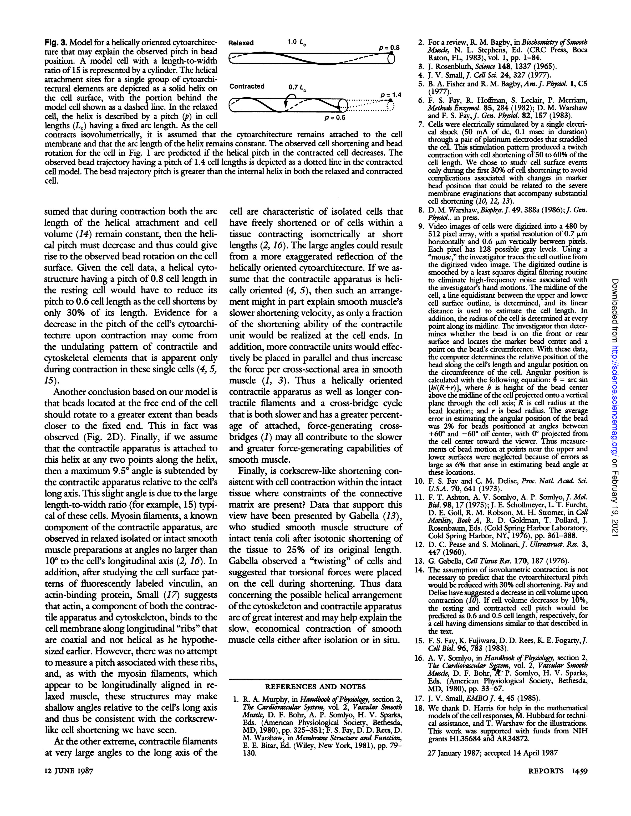
In addition to the contractile apparatus, the cytoskeleton must play an important role in the contractile function of a cell. Therefore, it is important to consider how the arrangement of the cell's cytoskeleton and contractile apparatus relates to the observed cell rotation upon shortening, reextension, and unwinding upon relaxation. One possible arrangement for these structures within the cell is in the form of a helix, as proposed by Small and Fisher and Bagby. Although it is not discernible from the bead rotational data whether the cytoskeleton or contractile apparatus is helically oriented within the cell, the bead data can be interpreted within the framework of at least one structural model (Fig. 3) in which the observed 1.4-cell length bead pitch is the result of a cytoarchitecture that is helically attached to the cell membrane.
If it is assumed that during contraction both the arc length of the helical attachment and cell volume remain constant, then the helical pitch must decrease, which could give rise to the observed bead rotation on the cell surface. Given the cell data, a helical cytostructure with a pitch of 0.8 cell length in the resting cell would need to reduce its pitch to 0.6 cell length as the cell shortens by only 30% of its length. Evidence for a decrease in the pitch of the cell's cytoarchitecture upon contraction may come from the undulating pattern of contractile and cytoskeletal elements that is apparent only during contraction in these single cells. Another conclusion based on our model is that beads located at the free end of the cell should rotate to a greater extent than beads closer to the fixed end. This was indeed observed (Fig. 2D).
Finally, if we assume that the contractile apparatus is attached to this helix at any two points along the helix, then a maximum 9.5° angle is subtended by the contractile apparatus relative to the cell's long axis. This slight angle is due to the large length-to-width ratio (for example, 15) typical of these cells. Myosin filaments, a known component of the contractile apparatus, are observed in relaxed isolated or intact smooth muscle preparations at angles no larger than 10° to the cell's longitudinal axis. Additionally, after studying the cell surface patterns of fluorescently labeled vinculin, an actin-binding protein, Small suggests that actin, a component of both the contractile apparatus and cytoskeleton, binds to the cell membrane along longitudinal "ribs" that are coaxial and not helical as he hypothesized earlier. However, no attempt was made to measure a pitch associated with these ribs, and, similar to myosin filaments, which appear longitudinally aligned in relaxed muscle, these structures may form shallow angles relative to the cell's long axis and thus be consistent with the corkscrew-like cell shortening we observed.
Conversely, contractile filaments at very large angles to the long axis of the cell are characteristic of isolated cells that have freely shortened or of cells within a tissue contracting isometrically at short lengths. The large angles could result from a more exaggerated reflection of the helically oriented cytoarchitecture. If we assume that the contractile apparatus is helically oriented, such an arrangement might partly explain smooth muscle's slower shortening velocity, as only a fraction of the contractile unit's shortening ability would be realized at the cell ends. In addition, more contractile units would effectively be placed in parallel, thereby increasing the force per cross-sectional area in smooth muscle. Thus, a helically oriented contractile apparatus, along with longer contractile filaments and a cross-bridge cycle that is both slower and has a greater percentage of attached, force-generating cross-bridges, may all contribute to the slower and greater force-generating capabilities of smooth muscle.
Finally, is corkscrew-like shortening consistent with cell contraction within intact tissue where constraints of the connective matrix are present? Data supporting this view have been presented by Gabella, who studied smooth muscle structure of intact tenia coli after isotonic shortening of the tissue to 25% of its original length. Gabella observed a "twisting" of cells and suggested that torsional forces were placed on the cell during shortening. Thus, data concerning the possible helical arrangement of the cytoskeleton and contractile apparatus are of great interest and may help explain the slow, economical contraction of smooth muscle cells either after isolation or in situ.
Figures (3)
Fig. 1. Successive digitized video images and corresponding computer-generated reconstructions. (A) A time series of digitized video images demonstrating corkscrew-like shortening and relaxation of a single smooth muscle cell attached only to a microprobe at its left end. The cell was decorated with marker heads. Beads are numbered and correspondingly labeled in (B) and (C). The time after stimulation is to the left of and below the cell. The cell length is presented above the cell as a percentage of the resting cell length. The curved arrows reflect the direction of rotation. Note that as the cell relaxed and reextended, rotation was opposite to that during shortening. (B) The same cell as in (A) after computer analysis of the digitized image. The computer analysis linearizes the cell. As in (A), the beads rotate about the cell during contraction. Closed circles represent beads on the front surface, whereas open circles are beads on the rear surface. (C) A view along the longitudinal axis of the cell in (B). As an example, beads 4 and 7 changed their angular bead positions during contraction, with bead 7 rotating to a greater extent than bead 4. (See above image for figure.)
Fig. 2. Graphs representing the contraction parameters for free shortening in smooth muscle cells. (A) Relative position of bead along cell length (L_r) as a function of percentage of cell shortening for the cell in Fig. 1. (B) Change in bead angular position on cell circumference as a function of time of contraction. The symbols and relative position of the bead as a percentage of L_c correspond to the beads in (A). Curves are the best fit to a third-order polynomial. The angular change for the bead at 85% L_c was not measured until 0.8 seconds after contraction because of its position on the top surface of the cell where errors in estimating bead angular position are large. (C) Change in bead angular position as a function of the percentage of cell length. The symbols and relative position of the bead (as a percentage) correspond to the beads in (A). From these data, a value for the pitch, describing the bead motion, was calculated by dividing the maximum slope determined from the polynomial fit by 360°. (D) The rate of normalized bead angular change as a function of the relative position of the bead beginning at the fixed end. Individual data points were obtained from the maximum slope for curves in (B) (filled circles). Triangles are data from another cell.
Fig. 3. Model for a helically oriented cytoarchitecture that may explain the observed pitch in bead position. A model cell with a length-to-width ratio of 15 is represented by a cylinder. The helical attachment sites for a single group of cytoarchitectural elements are depicted as a solid helix on the cell surface, with the portion behind the model cell shown as a dashed line. In the relaxed cell, the helix is described by a pitch (p) in cell lengths (L0) having a fixed arc length. As the cell contracts isovolumetrically, it is assumed that the cytoarchitecture remains attached to the cell membrane and that the arc length of the helix remains constant. The observed cell shortening and bead rotation for the cell in Fig. 1 are predicted if the helical pitch in the contracted cell decreases. The observed bead trajectory having a pitch of 1.4 cell lengths is depicted as a dotted line in the contracted cell model. The bead trajectory pitch is greater than the internal helix in both the relaxed and contracted cell.


